How Advanced Neuroimaging is Revolutionising TBI Diagnosis and Prognosis: A Game-Changer for Medico-Legal Cases
By Prof. Sotirios Bisdas, Consultant Neuroradiologist and MRI Lead
Posted 10 July 2025
6 Minute Read
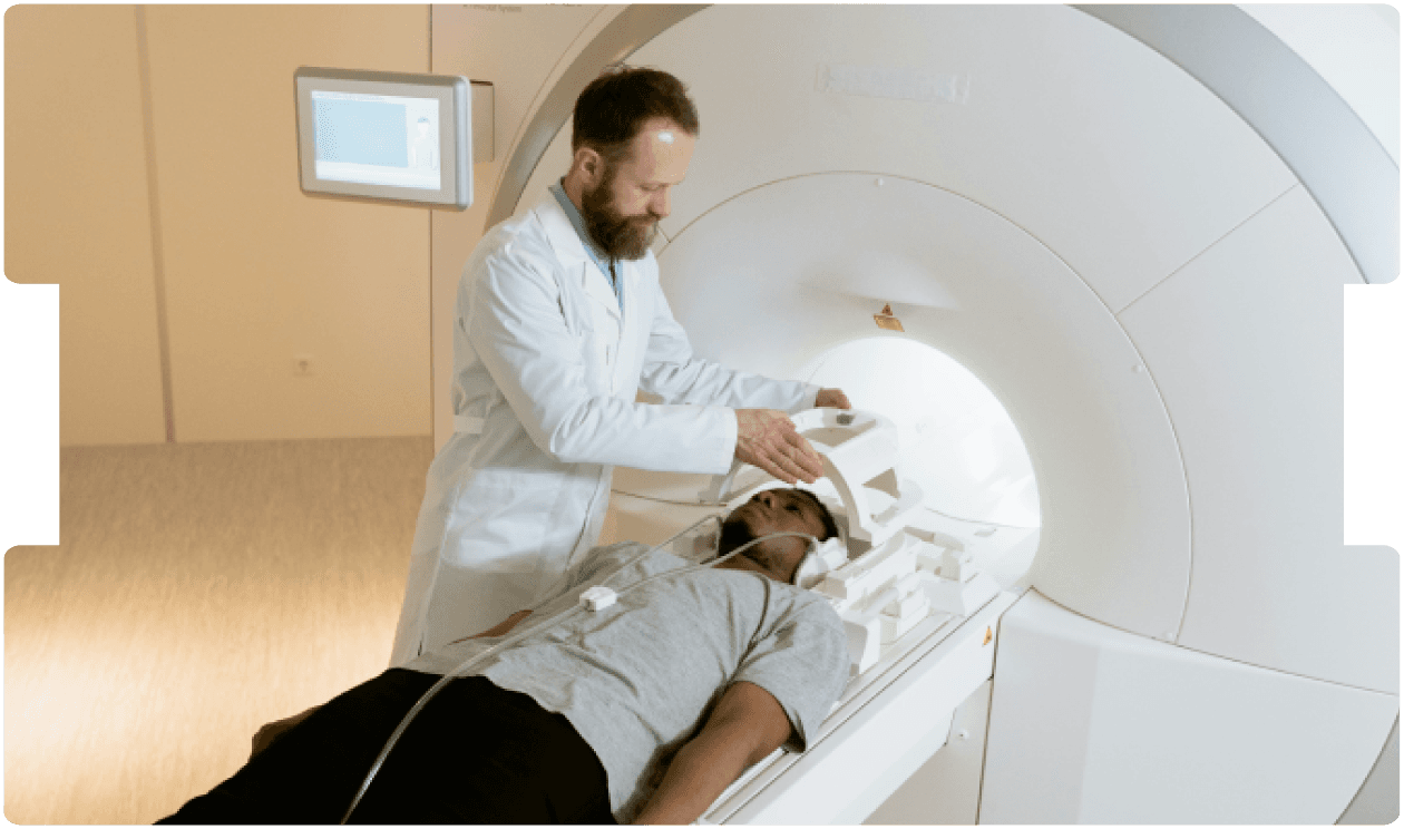
Revolutionary brain imaging is reshaping TBI claims - unlocking invisible injuries and empowering solicitors with robust, science-backed legal evidence.
Every year, millions of people worldwide suffer from traumatic brain injuries (TBI), ranging from mild concussions to severe trauma. While conventional CT and MRI scans have long been the standard for diagnosis, they often miss microscopic damage that can lead to debilitating symptoms such as chronic headaches, memory loss, mood swings, and cognitive impairments. For solicitors handling personal injury or medical negligence cases, this gap in detection can make it challenging to prove the extent of a client’s injury - until now.
The Limitations of Traditional Scans
Traditional imaging techniques like CT and MRI are effective at identifying bleeding, fractures, or large-scale tissue damage. However, they frequently fail to detect subtle white matter injuries or disruptions in neural networks - precisely the types of damage that can cause long-term cognitive and functional deficits. This leaves many TBI sufferers without a clear diagnosis, complicating their legal claims and access to appropriate compensation.
Cutting-Edge Neuroimaging: A Breakthrough for Legal Evidence
Advanced neuroimaging technologies, such as Diffusion Tensor Imaging (DTI) and resting-state functional MRI (rs-fMRI), are transforming how we diagnose and understand TBI. These tools provide unprecedented insights into brain injuries that were previously invisible, offering solicitors powerful evidence to support their clients’ cases.
1. Diffusion Tensor Imaging (DTI): Revealing Hidden Damage
DTI measures the movement of water molecules in the brain’s white matter - the "highways" that facilitate communication between brain regions.
In TBI, traumatic forces can cause:
· Axonal damage, disrupting critical neural connections
· Cell death and inflammation, altering water diffusion patterns
By quantifying these changes, DTI can identify microscopic injuries that correlate with symptoms like memory lapses, "brain fog," and balance issues. For solicitors, this means objective proof of injury even when traditional scans appear normal.
2. Resting-State fMRI: Mapping Functional Disruptions
While DTI shows structural damage, rs-fMRI reveals how different brain regions communicate.
TBI can lead to:
· Reduced connectivity, impairing cognitive functions
· Compensatory hyperactivity, where overworked circuits cause fatigue or mood disorders
· Network fragmentation, leading to inefficient processing
These functional disruptions often explain lingering symptoms misattributed to psychological causes. With rs-fMRI, solicitors can present concrete evidence of organic brain injury, strengthening claims for long-term care and compensation.
Who Can Benefit from Advanced Imaging?
These technologies are particularly valuable for:
· Clients with persistent symptoms but "normal" CT/MRI results particularly children whose injuries require specialist interpretation through paediatric neuroradiology.
· Suspected diffuse axonal injury (DAI), where DTI excels at detecting white matter damage
· Post-traumatic epilepsy cases, with rs-fMRI pinpointing seizure-related abnormalities
· Cognitive or behavioural impairments, helping tailor rehabilitation and legal arguments
Why This Matters for Legal Professionals
Advanced neuroimaging isn’t just a diagnostic tool - it’s a game-changer for building robust legal cases.
By providing:
· Clear, objective evidence of brain injury
· Correlation between injuries and symptoms
· Prognostic data to justify long-term care needs
Solicitors can secure stronger settlements and ensure clients receive the compensation they deserve.
The Future of TBI Claims
With studies showing that advanced MRI detects injury in 70% of TBI patients with normal conventional scans and predicts recovery with 85% accuracy, these technologies are setting a new standard in medical-legal evidence. For solicitors, staying informed about these advancements is crucial to advocating effectively for clients and holding insurers or defendants accountable.
Final Thoughts
For too long, TBI sufferers have faced scepticism due to the limitations of traditional scans. Now, advanced neuroimaging offers a scientific, irrefutable way to validate their injuries. As solicitors, leveraging these tools can make the difference between a dismissed claim and a life-changing settlement.
Is your client’s case being undermined by inconclusive scans? It might be time to explore advanced neuroimaging.
Tags:
- MRI Imaging
- Neurologist Expert Witness
- mTBI
- Missed Red Flags
- Missed Diagnosis
- Imaging Errors
- Brain Imaging Litigation
Expert Disciplines:
- Neuroradiology
About The Author

Prof. Sotirios Bisdas
Consultant Neuroradiologist and MRI Lead
Professor Sotirios Bisdas is a Consultant Neuroradiologist and Expert Witness with 12 years’ medico-legal reporting experience, having undertaken expert work since 2014. He has reported in over 80 cases across Clinical Negligence and Personal Injury, including breach of duty, causation and imaging evidence, with experience preparing joint statements and giving evidence at trial.
His specialist expertise includes neuro-oncology, traumatic and birth brain injury, hypoxic-ischaemic injury, stroke, spinal imaging, advanced MRI and PET imaging, making him particularly suited to complex neurological and neuroradiological instructions.
From the Blog
Related Articles
Sharpen your approach to complex claims involving functional symptoms. This expert-led guide helps distinguish genuine neurological disorder from malingering - crucial in high-stakes cases.

Spinal pain remains one of the most complex areas in medico-legal reporting, particularly where imaging does not align with symptom severity.

This blog guides clinical negligence solicitors on safely navigating virtual neurosurgery consultations - highlighting consent, standards, and documentation to strengthen medico-legal cases.
Find out why 70+ legal firms partner with INNEG.
Request a callback, or contact us.
INNEG respects your privacy. Any information you share with us will be used only to respond to your query.
Phone
+44 161 870 2461Thank you for your request!
We will get back to you as soon as possible.